Life Science Optics

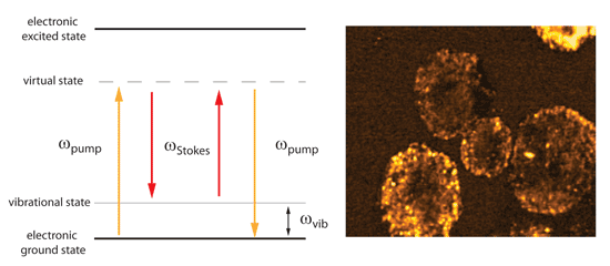
In SRS microscopy, like CARS microscopy, both the pump and Stokes photons are incident on the sample. If the frequency difference ωSRS = ωpump - ωStokes matches a molecular vibration (ωvib) stimulated excitation of the vibrational transition occurs. Unlike CARS, in SRS there is no signal at a wavelength that is different from the laser excitation wavelengths.
Instead, the intensity of the scattered light at the pump wavelength experiences a stimulated Raman loss (SRL), with the intensity of the scattered light at the Stokes wavelength experiencing a stimulated Raman gain (SRG). The key advantage of SRS
microscopy over CARS microscopy is that it provides background-free chemical imaging with improved image contrast, both of which are important for biomedical imaging applications where water represents the predominant source of nonresonant background
signal in the sample.
Figure 1: Stimulated Raman scattering (SRS) energy diagram for the SRS four-wave mixing process (right). Label-free stimulated Raman gain imaging of lipids in human melanocytes. Image courtesy of
Andreas Volkmer (University of Stuttgart, Germany).
Vibrational imaging based on stimulated Raman scattering microscopy
New Journal of Physics