
Stories & Features
Localization and quantitation of biochemical components in biological tissue are enabled through microscope-based detection of fluorescent probes that selectively bind to those components. Detection of the probe fluorescence is accomplished using an optical filter set, made up of one each exciter, dichroic, and emitter filter.
For single-probe detection, the exciter and emitter filters each have a single passband around the center wavelength and blocking outside the bandwidth. The dichroic has a long-pass edge near the wavelength at which the exciter and emitter filters’ optical transmission profiles cross over.
The long-wavelength spectral edge of the excitation filter and the short-wavelength edge of the emission filter play a critical role here and are called ‘critical edges’.
Many of the most useful fluorescent probes have small separations between their peak excitation and emission wavelengths. These so-called small Stokes Shift probes have presented a historical challenge to optical filter manufacturers, because the critical edges of the passbands could not be placed too close together, at risk of overlap due to filter manufacturing variability. The historical practice is shown schematically in Fig. 1, where the placement of the emission passband (blue trace) misses the prominent emission peak.
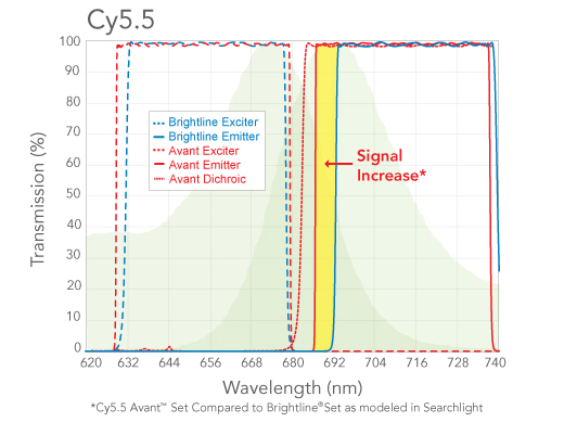
Fig. 1. Comparison of standard BrightLine® (blue) and Avant™ (red) passband positions for the Cy5.5 fluorophore. The yellow band highlights the fluorescence emission missed by the standard filter set.
In 2021, IDEX Health & Science devised the new Semrock Avant™ optical filter family to overcome this limitation. These sets were to improve upon the industry standard BrightLine® filter sets in three ways: (1) Improved control over manufacture to limit spectral edge variation in manufacture, (2) Steeper transition between transmission and blocking on the critical edges, and (3) Deeper complementary blocking adjacent to the critical edges.
This Case Study compares performance of filter sets made up of BrightLine and Avant bandpass filters, when used on a biological specimen.
A custom microscope slide was prepared by the Clemson Light Imaging Facility, Clemson University, Clemson, SC, in which the fluorescent probe Alexa Fluor 555 was used to stain the fast-twitch myosin isoform of sheep skeletal muscle. Images were acquired using a Nikon Plan Apo VC 20x/0.75NA objective lens, with a 5 ms exposure time, on a Nikon Ti2-E microscope system with a Photometrics Prime BSI camera, at the Center for Advanced Light Microscopy and Nanoscopy, University of Rochester Medical Center, Rochester, NY. The support by these facilities is gratefully acknowledged.
A comparison was made between the Avant filter set and the BrightLine filter set, both optimized for use with AF555. The imaging conditions were identical except for the filter set. Typical image results are shown in Fig. 2. The average signal increase
in the green pseudocolored regions was 20%, consistent with estimates of relative fluorescence emission made using SearchLight™, the simulator software developed by IDEX
Health & Science.
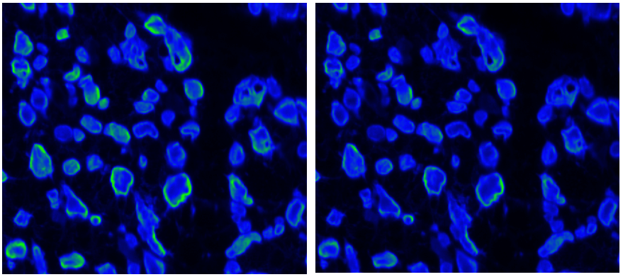
Fig. 2. AF555-stained myosin from sheep skeletal muscle imaged with Avant (left) and BrightLine (right) filter sets. See text for details.
This comparison validates the Avant filter set concept of using steeper edges with reduced edge jitter and increased blocking to increase signal and signal-to-noise in short Stokes Shift fluorescent probes.
Learn More About Avant Learn More About BrightLine Learn More About Semrock Optical Filters