
Stories & Features

IDEX Health & Science hosted our first Optics Photo Contest this past month. We hoped participants would join the life science optics community in the rare opportunity to feature their best scientific images curated with optical filters. We wanted to applaud individuals who exemplify mastery in their field and demonstrate their expertise through the use of state-of-the art optical filters to capture every photon possible.
To that end, we’re delighted to announce that, while we received many fantastic entries, we have officially selected our top two favorites as winners of the 2022 Optics Photo Contest. Each winner will be receiving a $50 VISA Gift Card and a customized YETI mug as a thank you for their participation.
Please join us in congratulating Daniel Han of Diatoms Australia and Daisuke Inoue of Kyushu University! Scroll down to see our favorite submissions. We hope to host another contest in the future, and we look forward to seeing the amazing scientific images your instruments are able to capture.
Check out our 2023 Optics Photo Contest
Learn more about our the filters that helped create these images
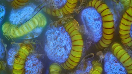
By: Daniel Han, Microscopist at Diatoms Australia
Image Title: Fern sori (spore capsule)
Image Details: “Spores in capsules found on fern leaves after a storm, autofluorescence. Ferns reproduce asexually and sexually. The asexual component known as sporophyte is commonly found. The autofluorescence is strong and beautiful with fern.”
Instrument or platform used to create this image: Olympus Microscope, Semrock DAPI filter set

By: Daisuke Inoue, Assistant Professor at Kyushu University
Image Title: Micropatterning of reconstituted microtubules
Image Details: “A stitched image of fluorescently labeled microtubules. In the image, microtubules were assembled on a micropatterned glass substrate.”
Instrument or platform used to create this image: Nikon Ti2-E inverted microscope (Nikon) equipped with a FITC Nikon fluorescence filter cube and a filter cube with a 50/50 mirror (Chroma). sCMOS Zyla5.5 (Andor) newly developed TIRF microscope (OPTO-LINE, Inc., Japan)