
Stories & Features
Silas Leavesley, Ph.D., is an associate professor of Chemical and Biomolecular Engineering and a member of the Center for Lung Biology at the University of South Alabama. His research focuses on the development of optical imaging and illumination technologies for use in novel biomedical and clinical imaging applications. Leavesley is co-founder of SpectraCyte, a startup business aimed at providing a next-generation gastrointestinal endoscope.
IDEX Health & Science: As a chemical engineer, you find ways to make existing products more efficiently. Your work, specifically, involves technologies for microscopy, endoscopy, and small-animal fluorescence imaging. Tell us more about this.
Silas Leavesley: The types of projects we work on look at the spectral nature of cells, tissues, or other biological samples. We use a technique called spectral imaging to extract more information from samples, and that information can be used to study what’s going on in a live cell or make a diagnosis about a tissue. Spectral imaging involves collecting image data at many different wavelengths, and there are different technologies you can implement to filter the image data at those wavelengths. We end up with what’s called a spectral image stack or spectral image cube. We take that information and run it through a range of different spectral imaging analysis algorithms, and we use the results to identify what molecules or components make up the tissue. If you’re looking at labeled molecules in the cell, for instance, you might be able to track those labeled molecules as they interact with one another. In a somewhat similar way, we can also look at native fluorescent molecules, or autofluorescent molecules, for tissue diagnosis. We’re also using spectral imaging to see if we can catch colon cancer at an earlier stage, to find things that are sometimes missed on colorectal exams. Those are some examples of how we are applying this technology.
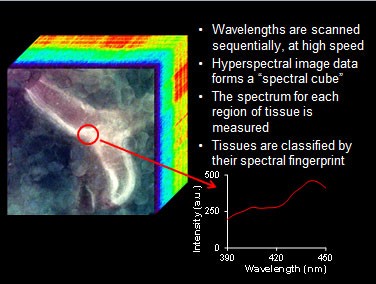
Principle of hyperspectral imaging of tissues. Image courtesy of Silas Leavesley, University of South Alabama.
IDEX Health & Science: What drew you to this type of work?
Silas Leavesley: I’ve always wanted to do work that has a real impact on our ability to diagnose or treat patients. This area of research is one way that we can improve our ability to diagnose and then ideally to treat cancer, by developing new devices to diagnose it earlier or stage it more accurately. I also enjoy imaging, so I’ve been very interested in the ability to take imaging and couple it with the spectroscopic component. One other thing that’s really drawn me to this work is that I enjoy being able to design and implement new technologies. We can come up with new types of detection modalities or imaging modalities, envision how a final product might look, and figure out what specifications we need to develop it.
IDEX Health & Science: You developed, along with Dr. Thomas Rich, hyperspectral imaging fluorescence microscopy technologies that improve detection specificity and sensitivity for clinical diagnostic devices. Can you talk more about this technology and your startup, SpectraCyte?
Silas Leavesley: The goal of this company is to take what we do in the lab and transition it to more end-user devices. How do we take concepts like spectral imaging and envision it as a final product that can detect colorectal cancers at an earlier stage, and then how do we implement that final device and get it to the clinician? We tried to go through all the steps in that process, and what we found on the research side is that you can only go so far before it’s difficult to get funded by the standard research grant mechanisms. So to really start developing translational devices, we decided to see if we could do this as a startup company and get funding that supports the development of devices that we can start testing for clinical trials. We’ve applied the spectral imaging technology on our microscope platform; we’re also working on a prototype endoscope that can operate at very high speeds so we can perform spectral imaging in real time. We’re working on getting the endoscope into the clinic, and then we would ideally branch off and look at surgical devices like laparoscopes and possibly other scopes.
IDEX Health & Science: You’ve developed technologies and methods using Semrock’s VersaChrome® tunable filters that continue to be at the leading edge in the scientific field. Can you tell me more about how Semrock products have helped you in your work?
Silas Leavesley: On the microscope side, we were able to work with Semrock to come up with a system that allowed us to sample spectral image data on a range of wavelengths: UV up to red wavelengths. At the same time, we can acquire a strong signal. The combination of those helped us be able to perform spectral imaging for really high quality images of cells and tissue, and we’ve applied that in a couple of different ways. One way was using the microscope system to show how we can detect specific molecules like fluorescent proteins or other labels; maybe you’re labeling one cell in a tissue or one specific component of a cell, and you can pull that signal out in the midst of a very high background that’s called tissue autofluorescence. We’ve also taken that technology and used it in some of our preliminary testing for characterizing tissues. We had an article come out recently in the Journal of Biomedical Optics where we looked at an initial sample set of colorectal cancer tissues, screening them with the VersaChrome filters to show proof of principle that the spectral properties of cancerous tissues look different than the spectral properties of normal surrounding tissue.
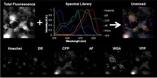
Multilabel assessment of cellular compartments. Multiple, overlapping labels can be used in live-cell assays. Hyperspectral imaging can be used to localizing signals to multiple subcellular compartments. Image courtesy of Silas Leavesley, University of South Alabama.
IDEX Health & Science: Overall, how has the field of cytometry advanced since you got started?
Silas Leavesley: When I first got involved in this field, about a decade ago, the idea of using a technique like spectral imaging in microscopy had already been demonstrated in several ways, but the applicability was limited by the technology available and how easy those technologies were to apply to a biological problem. One of our focuses has been to develop new implementations of technology to improve the capabilities of spectral imaging in microscopy with regard to signal strength, speed, and quality, but then also to educate and raise awareness about what these technologies can do in different settings and different application areas. One area that we’ve focused on has been second-messenger signaling; we’ve been trying to show what we can do with spectral imaging in microscopy—how to keep pushing the boundaries for acquiring image data of second-messenger signals in discrete compartments within the cell and how the signal may help to dictate the overall physiological response of the cell. The field of compartmentalized cell signaling is evolving and the need for detecting subcellularly-localized signals is growing. We’ve tried to help develop approaches that make spectral imaging technologies easier to implement and more straightforward.
IDEX Health & Science: You have been actively involved in the Cyto congress (ISAC) in the past. What is unique about this conference compared to other scientific meetings?
Silas Leavesley: Cyto to me is an outstanding conference because it brings together a mix of people. Cyto developed out of the flow cytometry field, so a large focus of the conference remains on flow cytometry. Flow cytometrists have always also been, to a large part, inventors. The conference is a mix of clinicians, flow cytometrists, and imaging people, and all of them bring some aspect of innovation or technology development. I run into a range of people that work on the technology side but also on the application side and the clinical side, and it’s a really neat environment for all those people to get together and dialogue. They are very good people to work with at an interdisciplinary level.
IDEX Health & Science: What would you consider your biggest achievement to date?
Silas Leavesley: We’re pushing a new type of spectral imaging technology called excitation scanning spectral imaging. I think our biggest achievement to date is getting the prototype system out on both the microscope and now on the endoscope that use that technology. We’ve gotten to the point where we’ve implemented it, we’re now evaluating it, and I think it’s going to be out there as an end-user type system within the next couple of years.