Multiband Filter Set Terminology
The ability to label multiple distinct objects of interest in a single sample is an important asset to fluorescence imaging. For a long time the only way to achieve high-quality images of such samples was to take multiple photographs while switching whole single-band filter cubes between photographs and then later combine these photographs electronically. Limitations to this approach included pixel shift among the multiple, single-color images and the speed with which a complete multi-color image could be captured.
While the problem of pixel shift can be eliminated with Semrock's BrightLine ZERO filter sets, the single-band filter cube approach, although slow, remains the best technique for achieving images with the highest contrast and lowest bleedthrough. To satisfy the ever-increasing demand for high-speed imaging, especially for live-cell real-time analysis using fluorescent protein labels, there is a need for an alternative to the single-band filter cube approach without sacrificing image fidelity. Recent advances in multiband optical filter technology have brought simultaneous multi-color imaging to a new level.
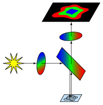
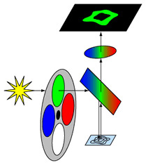
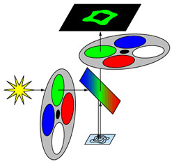
There are three types of multiband filter sets for simultaneous multi-color imaging. The "full-multiband" configuration uses a multiband exciter, emitter, and dichroic beamsplitter and is ideal for direct visualization, such as when locating areas of interest on a sample. This approach is quick and easy to implement and is compatible with all standard fluorescence microscopes, though it requires a color camera for electronic imaging and cannot eliminate fluorophore bleedthrough. The "Pinkel"configuration uses single-band exciters in a filter wheel with multiband emitter and dichroic filters, and offers an economical way to achieve very high-speed, high-contrast, simultaneous multi-color imaging. This approach is based on a monochrome CCD camera, which is less expensive and offers better noise performance than color cameras. While bleedthrough is reduced relative to the full-multiband approach, some bleedthrough is still possible since all emission bands are imaged simultaneously. The "Sedat" configuration uses single-band exciters and single-band emitters in synchronized filter wheels, with a multiband dichroic beamsplitter. This approach provides the best image fidelity for high-speed simultaneous multi-color imaging, though it requires a larger investment in system hardware.
"Full Multiband" Configuration (Multiband exciter, multiband emitter, & multiband dichroic)
"Pinkel" Configuration (Multiband emitter, multiband dichroic, & single-band exciters)
"Sedat" Configuration (Multiband dichroic, single-band exciters, & single-band emitters)
"Full Multiband" Image Multicolor image captured with a color CCD camera
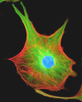
Image of bovine pulmonary artery endothelial cells (Molecular Probes FluoCells #2 reference standard)
"Pinkel" or "Sedat" Composite Image Single-color images are combined electronically to produce one high-fidelity, multi-color image.
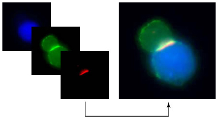
T-Cell and Antigen Presenting Cell (APC) conjugates demonstrating an immunologic synapse. Samples courtesy Beth Graf and Dr. Jim Miller at the University of Rochester Medical Center.
Learn More About Multiband Fluorescent Imaging:
ReadNew Optical Filters Improve High-Speed Multicolor Fluorescence Imaging from the March 2006 issue of BioPhotonics