Laser-based Imaging: What you need to know
Optical Filters for Laser-based Fluorescence Microscopes
Introduction
Due to many desirable properties like high brightness, stability, longevity and narrow spectral bandwidth, lasers are replacing conventional broadband light sources for fluorescence imaging applications. Not only have these features of lasers allowed for higher-sensitivity visualization and enhanced throughput in imaging applications, but also several other unique properties of lasers—narrow beam divergence, high degree of spatial and temporal coherence, and well-defined polarization properties—have enabled new fluorescence imaging techniques. At the same time, the use of lasers as fluorescence light sources imposes new constraints on imaging systems and their components. For example, optical filters used in laser-based imaging systems such as confocal and Total Internal Reflection Fluorescence (TIRF) microscopes have unique requirements compared to those filters used in broadband light source based instruments.
Optical Filters Optimized for Lasers
Various types of lasers have evolved over the last four decades. Lasers tend to be classified by the gain medium and pumping scheme. Until recently the most popular lasers for fluorescence imaging have been gas lasers, such as Ar-ion and Kr-ion
lasers. More recently, solid-state lasers have been replacing gas lasers. Popular laser types include semiconductor diode lasers, optically pumped semiconductor lasers and frequency-doubled diode-pumped solid-state (DPSS) lasers. Table 1 summarizes
the most popular lasers used for fluorescence imaging applications.
Excitation Filters: Contrary to popular opinion, optical source clean-up filters (excitation filters) are important for laser sources to block the unwanted light at wavelengths away from the actual laser line, including spontaneous emission observed in solid-state lasers and the plasma lines of gas lasers. Not only do these filters provide deep blocking of the unwanted light (typically OD > 6), but they also provide exceptional transmission at the laser lines (see Figure 1). Additionally, these filters should be durable enough to withstand the high intensity of laser beams. Unlike the traditional soft-coated fluorescence filters used for decades, newer hard-coated thin-film filters have a much high laser damage threshold (LDT) ratings. For continuous wave (cw) lasers, this LDT rating corresponds to about 10 to 100 kW/cm2. High optical durability, combined with the environmental reliability of hard-coated filters eliminates the need to ever replace the filters for most fluorescence microscopy applications.
Table 1: Lasers for popular fluorophores. "DPSS" = diode-pumped solid-state laser. "OPS" = optically pumped semiconductor laser. "Doubled" = frequency doubled via a nonlinear optical crystal.
| Laser Line | Laser Type | Popular Fluorophores |
|---|---|---|
| ~ 405 nm | Diode | DAPI, Hoechst, Alexa Fluor 405TM, BFP |
| ~ 440 nm | Diode | CFP |
| 473 nm | Doubled DPSS | |
| 488.0 nm | Ar-ion gas | GFP, FITC, Alexa Fluor 488TM |
| ~ 488 nm | Doubled OPS | |
| 514.5 nm | Ar-ion gas | YFP, Rhodamine |
| 515.0 nm | Doubled DPSS | |
| 561.4 nm | Doubled DPSS | TRITC, Cy3TM, RFP mCherry |
| 568.2 nm | Kr-ion gas | |
| 593.5 nm | Doubled DPSS | Texas Red, mCherry (mRFP) |
| 632.8 nm | HeNe gas | |
| ~ 635 nm | Diode | Cy5TM, Alexa Fluor 647TM |
| 647.1 nm | Kr-ion gas |
The spectral characteristics of these excitation filters are dictated by the type of laser. Since gas lasers and DPSS lasers, have very precise and narrow laser lines the ideal excitation filter is a narrowband laser-line filter (typical bandwidth < 0.4% of the laser wavelength), keyed to the precise location of the laser line. These narrow laser line filters are not, however, a good match for systems that might use multiple lasers with similar wavelengths or for semiconductor lasers. The spectral output from diode and optically pumped semiconductor lasers can vary appreciably from laser to laser, with temperature, and as the lasers age. For these lasers broader excitation filters that appear similar to those used for broadband light source microscopy systems are the better choice. For example, the excitation filter shown in Figure 1 is designed to be used with both 375 and 405 nm lasers, with the long-wavelength edge taking into account a ± 5 nm uncertainty in the wavelength of the 405 nm laser. While similar, these excitation filters are not identical to broadband light source filters. In addition to edge positions of laser filters being precisely keyed to the associated laser wavelengths, edge steepness as well as ripples in the passband are other important considerations. Low ripple ensures high transmission at specific laser lines or over time as a semiconductor laser wavelength drifts and steep edges provide high optical noise discrimination.
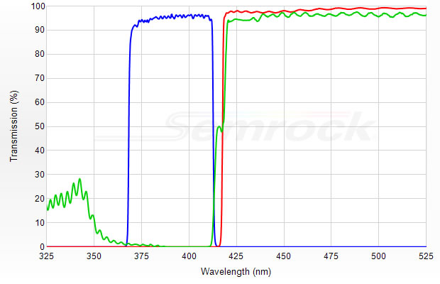
Figure 1: Measured spectral performance of a typical laser fluorescence filter set; blue line – exciter; green line – dichroic; red line – emitter.
Emission Filters: The emission filter should provide high blocking (> OD 6) at all laser lines that might be used with the filter set, thus ensuring the darkest background signal level, while at the same time providing very high (> 97%) transmission of the emission signal. Emission filters for broadband light sources generally do not provide sufficient blocking at laser lines, leading to an appreciable compromise in imaging contrast. As with the excitation filter, the edge wavelength of an emission filter should be precisely keyed to block the associated laser lines, and the edge steepness of the short-wavelength edge is especially critical. Additional considerations for emission filters include the use of high optical quality glass for the substrate that exhibits low autofluorescence, excellent homogeneity, and low wedge angle for minimal beam deviation that can lead to pixel shift when exchanging filters.
Demanding applications such as imaging of single molecules, ideally suited for laser excitation, may impose additional constraints on the blocking of laser beams in the emission channel while maximizing the collection of every possible photon from the
fluorophores. In such situations, conventional bandpass emission filters may be replaced by long-wave-pass filters (Fig. 1). Long-wave-pass emission filters also allow capture of maximum signal from fluorophores that have widely separated absorption
and emission spectra. In our experience, some demanding applications (especially TIRF systems) even benefit from using a second emission filter or a notch filter in conjunction with all the filters of a laser set. The main purpose of the second
filter, which should be physically separated from the first emission filter, is to ensure that higher-angle scattered excitation light does not make it through the entire imaging path to the detector.
Dichroic beamsplitters: Dichroics for laser applications are designed with their reflection and transmission bands matched to the excitation and emission filters and have antireflection coatings to maximize transmission of the emission
signal and eliminate coherent interference artifacts. Dichroics also have similar LDT ratings to those of the excitation filters and should have low ripple in the reflection bands to minimize variation of the excitation intensity.
Since the dichroic beamsplitter is directly exposed to the powerful excitation beam, even weak autofluorescence from the filter will contaminate the emission signal. Therefore, a substrate with ultra-low autofluorescence, such as fused silica, should
be used. Note that since the excitation light and the emission signal intensity levels differ by many orders of magnitude (typically a factor of 106) the requirement for the emission filter autofluorescence is not as stringent as for the dichroic
beamsplitter.
The flatness of the dichroic has significant impact on image quality. For most laser microscope applications, the dichroic should be flat enough such that there is no noticeable shift in the focal spot of the illumination laser beam, where focal shift
is typically defined by the Rayleigh range. This is critical for applications such as TIRF microscopy and structured illumination.
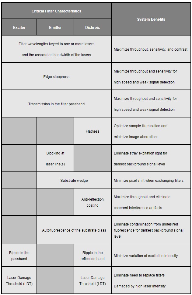
Optical filters working together as a set: Table 2 includes a summary of the critical characteristics of filters for laser systems. Overall, it is desirable that the optical filters achieve high blocking as well as high transmission of specific wavelengths of light without compromising the diffraction-limited image quality. This simple set of requirements not only influences the design of an individual filter but of the system of filters that are used in combination. Therefore, the designs of the excitation and emission filters as well as that of the dichroic beamsplitter should be complimentary to obtain the highest fidelity fluorescence visualization.
Table 2: Summary of critical characteristics of optical filters specifically for laser imaging systems.