Introduction to Fluorescence Filters

Fluorescence occurs when a molecule absorbs light at wavelengths within its absorption band, and then emits light at longer wavelengths within its emission band. For example, brightly fluorescent molecules (called fluorophores) can be attached to biologically significant molecules in e.g. cell membranes, in the brain, or even on subunits of DNA, whose structures become visible in a fluorescence microscope that allows us to track the way cells function in health and disease. Fluorescence is widely used in biology, biotechnology, and medicine, due to its extraordinary sensitivity, high specificity, and simplicity of usage.
For a more in-depth overview of considerations relevant to the design of a fluorescence multiplexing system, click here.
What Optical Filters are Included in a Fluorescence Instrument?
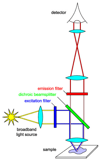
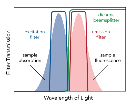
Fluorescence microscopy, and most instruments that use fluorescence, including fluorescence microscopes, rely on optical filters. A typical microscope has three basic filters: an excitation filter (or exciter), a dichroic beamsplitter (or dichroic mirror), and an emission filter (or emitter). These three filters form what is referred to as a “filter set” and are often housed in a special assembly called a “filter cube” that can be quickly mounted in a microscope.
The excitation filter is usually a bandpass filter designed to pass only those light source wavelengths that are to be absorbed by the fluorophore, thereby minimizing excitation of other sources of fluorescence, and especially blocking excitation light in the fluorescence emission band.
The dichroic is an edge filter (usually passing longer wavelength light and reflecting shorter wavelength light) used at an oblique angle of incidence (typically 45°) to efficiently redirect light in the excitation band towards the sample and transmit light in the emission band towards the detector.
The emitter is usually a bandpass filter that passes only the wavelengths emitted by the fluorophore and blocks all undesired light outside this band – especially the excitation light.
The main difference between an excitation filter vs emission filter? An excitation filter lets in light to excite a molecule, while an emission filter transmits the light the molecule emits.
An optimal optical filter set of the above three individual filters blocks unwanted excitation light (including in the UV and IR) as well as unwanted emitted light (including autofluorescence) to ensure high signal and low background.
The filter set described above can only be used for one fluorophore, since it has only one passband each for excitation and for emission. If for example two fluorophores are used, the set will need to have two passbands each for exciter, dichroic, and emitter. These are called multiband sets; for more information, review our White Paper.
What are important characteristics of filter sets?
The type of experiment being performed often determines which type of filter set should be used in the microscope:
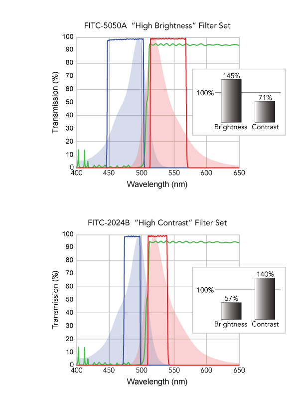
- High Brightness: This type maximizes the detected signal, even at the expense of higher background.
- High Contrast: This type maximizes image contrast between bright areas and background, i.e., for high signal-to-background ratios.
- Narrow passbands: When using multiple fluorophores, reducing crosstalk, and/or decreasing background signal, narrow passbands ensure only the desired signal from the desired fluorophore is captured.
- Wide passbands: When using a weakly emitting fluorophore or imaging single molecules, wider passbands allow more excitation light to the sample and capture more emission.
To summarize, filter sets strike a balance between high brightness and high contrast, typically by using narrower or wider passbands in combination with center wavelength placement.
How Can Semrock Optical Filters and Filter Sets Optimize Your Fluorescence Application?
The team at IDEX Health & Science has specific experience and expertise in design of optical filter sets and optimizes each set to make sure the filters work well together. Semrock fluorescence optical filters and fluorescence filter sets therefore offer a number of critical advantages to users and developers of fluorescence-based optical platforms/p>
- Semrock filters are based on manufacturable specifications, which guarantee excellent filter-to-filter and instrument-to-instrument spectral consistency. Learn more.
- Semrock measures the spectral performance of production filters to the highest fidelity, using proprietary instruments that very accurately measure steep spectral edges and deep OD, and ensure that the produced spectral performance meets or exceeds the specified performance. These instruments include the workhorse Peregrine platform that overcomes the high spectral resolution and sensitivity limitations of standard metrology equipment, and KolaDeep™ that routinely measures blocking down to beyond OD9 and edge steepness steeper than 0.2% relative to the edge wavelength. These allow Semrock to deliver assured performance in the most demanding of situations, meeting the needs of a new generation of precision fluorescence platforms. Learn more.
- Semrock can coat catalog and custom optical filters with Precision Edge Placement, which delivers industry-low manufacturing tolerances for critical filter spectral edges.
Semrock employs all these advantages in the industry-leading Avant™ filter sets that increase emission signal from short Stokes’ shift (low “Gap”) fluorophores by collecting the emission signal over the peak emission ranges and blocking excitation crosstalk. Learn more.
Our flagship BrightLine® filter sets follow fluorophore usage trends, light source innovation, and detector enhancements. These sets balance filter set needs and cost and offer set performance over a wide range of fluorescent probes and microscopy applications, based on our years of experience designing optical filters.
What is a real-life example of using fluorescence filter sets?
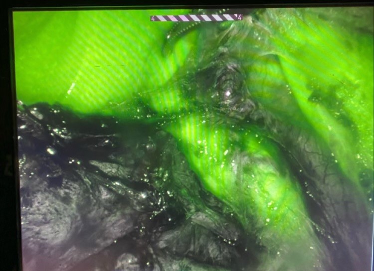
Fluorescence guided surgery (FGS) uses optical filters to assist surgeons in visualizing organs or structures during surgery. In this example, the patient drank a fluid containing the fluorophore, and the surgeon used a special visualizing system that sent light into the body to excite the fluorophore. In the photo, the green area highlights an anatomically complex location, and the surgeon knows precisely where to operate. Without FGS, this patient’s surgery would be difficult and dangerous.
Learn More about Fluorescence Guided Surgery
Photo Credit: Brianna Nelson
Resources
How do I know what filter set I should use?
We have tools to assist in selecting the filter set that matches your needs:
- Looking for a filter set to go with your fluorophore? Visit our reference chart.
- Use our SearchLight™ tool to evaluate the spectral performance of your fluorescence microscopy set-up.
- Have questions about optical filters for fluorescence applications? Check out our FAQ page.
- Selecting Filters for Fluorescence Multiplexing (White Paper)
- Visit our Semrock Resource Page for more information.
How do I install a filter set in a filter cube?
Explore our guide for many common filter cubes.