Choosing Dichroic Beamsplitters with Flatness/RWE Appropriate to the Microscopy Method
 Wavefront
distortion can degrade image quality by reducing contrast or compromising resolution. In several microscopy applications, reducing wavefront distortion is critical to achieving the microscopy method. Specifying and selecting optical filters that minimize
wavefront aberration is important to maximize or enable optical system performance. This Tech Note elucidates how to select optical filters for high performance microscopy, and provides guidance on choosing Semrock brand catalog filters for wavefront
distortion performance required for applications.
Wavefront
distortion can degrade image quality by reducing contrast or compromising resolution. In several microscopy applications, reducing wavefront distortion is critical to achieving the microscopy method. Specifying and selecting optical filters that minimize
wavefront aberration is important to maximize or enable optical system performance. This Tech Note elucidates how to select optical filters for high performance microscopy, and provides guidance on choosing Semrock brand catalog filters for wavefront
distortion performance required for applications.
Both standard and advanced microscopy methods require certain minimum standards of flatness on dichroic beamsplitters. For example, super-resolution and TIRF microscopy cannot be achieved if the flatness of the critical dichroic beamsplitters is worse than required. This Tech Note introduces the topic of how to choose Semrock dichroic beamsplitters appropriate to the microscopy method.
Optical filters are generally composed of multi-layered, thin-film coatings on plane, parallel glass substrates. There is variability in substrate flatness, and, additionally, the substrate may slightly bend after coating. For transmitted light, such bending has little effect on transmitted wavefront error (TWE) other than a slight displacement of the beam axis. However, for reflected light, especially light incident at non-perpendicular angles, deviations from flatness (see Figure 1) have two effects on reflected wavefront error (RWE): (1) the focal plane may shift position, or (2) the beam may acquire optical aberrations (e.g., astigmatism).
(1) Radius of Curvature (m) ~6
(2) ~1275
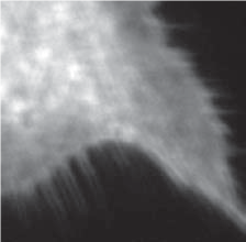
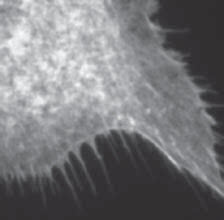
A focal plane shift can be corrected by lens or camera adjustment in some microscopy methods, but in others this shift cannot be adjusted for and can therefore impede proper function. Also of significant concern, optical aberrations can degrade image quality, as illustrated in figure, image sequence demonstrating the effect of substrate bending on image quality. A lower radius of curvature means greater substrate bending. As the radius of curvature decreases (and bending increases), image quality degrades. [Images o F-actin in bovine pulmonary artery endothelial cells (FluoCells® Prepared Slide #1,ThermoFisher Scientific, Waltham, MA, USA) as imaged on a BX41 microscope (Olympus Corporation of the Americas, Center Valley, PA, USA) with a 40×, 0.75NA objective and Retiga camera (QImaging, Surrey, BC, Canada)]
A focal plane shift can be corrected by lens or camera adjustment in some microscopy methods, but in others this shift cannot be adjusted for and can therefore impede proper function. Also of significant concern, optical aberrations can degrade image quality, as illustrated in above Figure, and can compromise the microscopy method. In determining suitable flatness need for a given application, the most important parameter is often the diameter of the beam striking the dichroic beamsplitter surface. Reference the table in the "What is flatness / RWE classification?" FAQ to find the Semrock optical filters best suited to the application, for maximum diameter values. For more specific information, and for other beam diameter value and microscopy examples, the white paper on this topic [1] provides additional information on RWE, TWE, and microscopy methods, as well as guidance from a system designer’s perspective.
Because of potential degradation of imaging quality, it is important to determine when the flatness of dichroic beamsplitter is critical to a microscopy method. Table 1 lists examples of popular microscopy techniques in which it is critical to have dichroics of high flatness. As noted in this table, high flatness requirements can apply to both the illumination (excitation) and the detection (emission) light paths.
Table 1: A list of popular standard and advanced microscopy methods, categorized as to criticality of dependence on reflected wavefront flatness.
Microscopy Technique | Flatness Need in a Reflected Excitation Beam | Flatness Need in a Reflected Emission Beam |
|---|---|---|
Widefield Fluorescence Microscopy | Non-critical | Critical |
Total Internal Reflection Fluorescence (TIRF) Microscopy | Critical | Critical |
Stochastic Switching (PALM, STORM, etc.) | Critical | Critical |
Stimulated Emission Depletion (STED) – Pulsed Microscopy | Critical | Non-critical |
Confocal Single-point Scanning Microscopy | Critical | Non-critical |
Combining multiple laser beams | Critical | Not Applicable |
IDEX Health & Science offers an extensive and industry-leading range of Semrock brand catalog filters for a variety of applications with specific Flatness / RWE needs. The Flatness Classifications listed in the "What is flatness / RWE classification?" FAQ provides an intuitive approach to selecting products of appropriate flatness for a given application. For more specific information, and for other beam diameter value and microscopy examples, the white paper on this topic [1] provides additional information on RWE, TWE, and microscopy methods, as well as guidance from a system designer’s perspective.